Rib Cage Muscles Anatomy : Human rib cage, 3/4 front view | Skeleton anatomy, Human ... / But for an anatomy study, it's not.. Related posts of muscle anatomy rib cage anatomy muscle cell. Structure of a typical rib: They are each attached to the ribs. We study anatomy at the practical anatomy class we study the human body. Muscle spasms located in the rib cage are often observed in people who strain or overwork their upper body muscles.
Rib cage anatomy and breathing. Abdomen & ribs muscle movements. The muscular system consists of the skeletal muscles and their associated structures. The rib cage, shaped in a mild cone shape and more flexible than most bone sets, is made up of varying elements such as the thoracic vertebra, 12 equally paired ribs, costal cartilage, and held together anteriorly by the sternum. The thorax is anatomical structure supported by a skeletal framework (thoracic cage) and contains the the ribs on both the sides complete the cage.
Learn Muscle Anatomy: Serratus Posterior Superior and Inferior from cdn2.hubspot.net They are each attached to the ribs. The ribs joint as follows muscles of the thoracic wall contain those that fill and support the intercostal spaces, those that pass between the sternum and the ribs, and those that cross several. The intercostal muscles are the muscles that occupy the 11 intercostal spaces. Ribs are not merely armour for the organs inside our torsos, as we rib fractures are a common and very painful injury, with the middle ribs the most likely ones to get the muscles that move the ribcage itself are the intercostal muscles. Print of thorax ribs cage anatomy illustration 1866. For a gesture drawing, that's good enough. Injuries to your rib cage or muscles in your upper chest can cause rib pain ranging from a dull ache to sudden, sharp jabbing pains in the affected area. See more ideas about anatomy, anatomy study, rib cage anatomy.
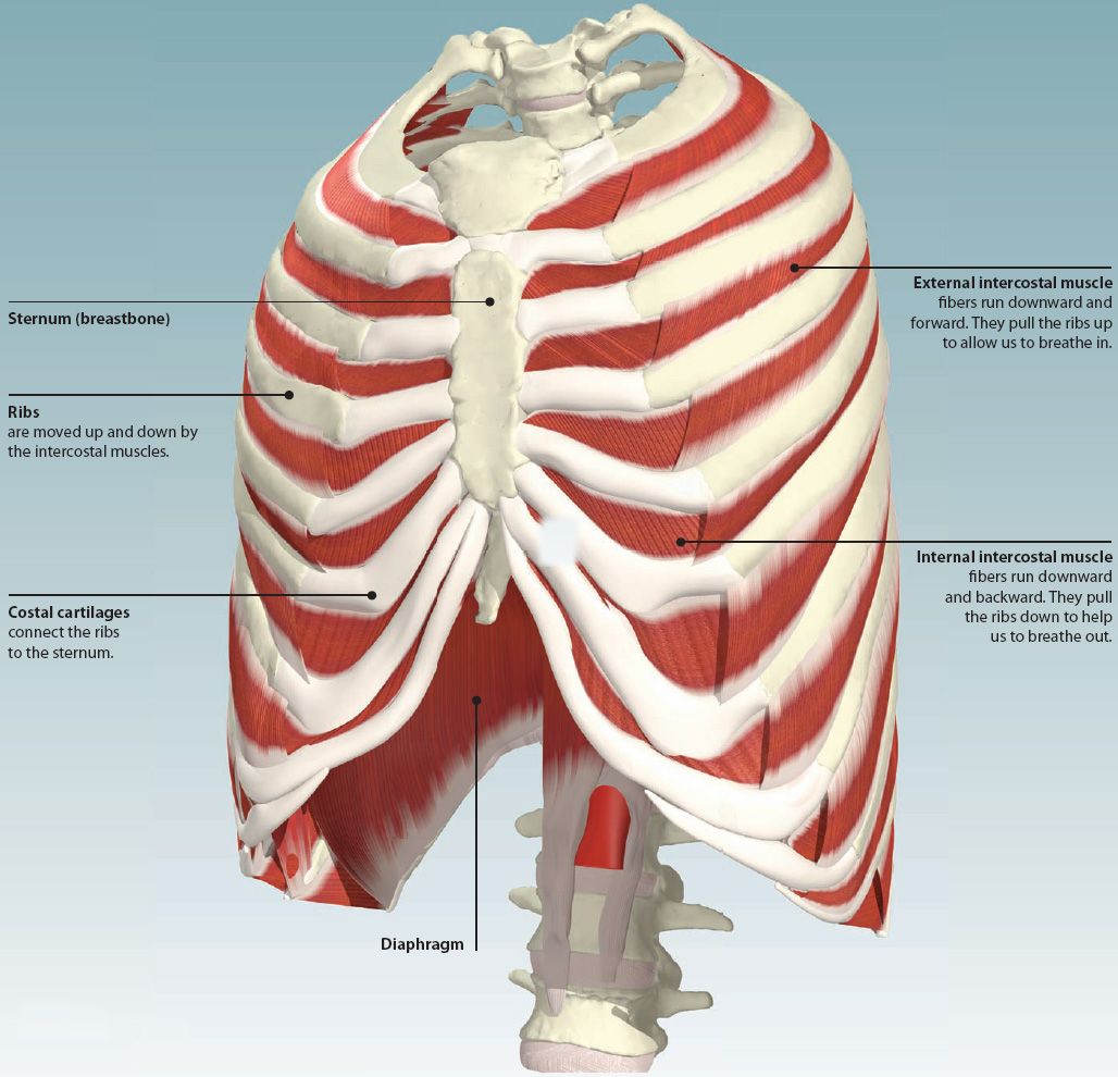
Structure of a typical rib:
The rib cage is composed by sternum, costal cartilages, and ribs connected to the thoracic vertebrae. The back end is wide and open. But for an anatomy study, it's not. During normal breathing, contraction of the major inspiratory muscle, the diaphragm, produces both rib cage expansion and a downward movement of the diaphragm. They are more involved in forced expiration and coughing to forcibly shrink the chest and. Anterior view of the lungs and ribcage in a transparent female torso stock illustration these pictures of this page are about:human anatomy rib cage muscles. Your rib cage plays an important role in respiration, expanding and contracting as your respiratory muscles, including your diaphragm, work to help you breathe. Skeletal muscles attached to the rib cage: This is a stereogram, to be viewed in crossview technique. The ribs form the main structure of the thoracic cage protecting the thoracic organs, however their main function is to aid respiration. The rib cage is often simplified as an oval shape. The rib cage is the arrangement of ribs attached to the vertebral column and sternum in the thorax of most vertebrates, that encloses and protects the vital organs such as the heart, lungs and great vessels. Muscle spasms located in the rib cage are often observed in people who strain or overwork their upper body muscles.
Try to be as accurate as you can with them. Ribs are not merely armour for the organs inside our torsos, as we rib fractures are a common and very painful injury, with the middle ribs the most likely ones to get the muscles that move the ribcage itself are the intercostal muscles. The following general rules regarding actions can be. Serratus posterior superior and inferior. Print of thorax ribs cage anatomy illustration 1866.
Image result for Rib | Human body anatomy, Anatomy bones ... from i.pinimg.com The other attachment of these muscles is usually considered to be either superior or inferior to the rib attachment. Skeleton drawings anatomy for artists anatomy reference. The rib cage is a primarily protective structure, encircling the heart and lungs. This cage protects vital organs and is essential for creating negative pressure to inflate lungs. The rib cage is often simplified as an oval shape. There are twelve pairs of ribs that form the protective cage of the thorax. 876x1024 bony walls of the thorax clipart etc. For a gesture drawing, that's good enough.
This is a stereogram, to be viewed in crossview technique.
Illustration of rib cage, demonstrating ribs and connection through cartilage to sternum. But for an anatomy study, it's not. But the cartilages of these ribs are not. The ribs form the main structure of the thoracic cage protecting the thoracic organs, however their main function is to aid respiration. Muscles that move the rib cage attach to the rib cage. The rib cage is a primarily protective structure, encircling the heart and lungs. The ribcage is made to be flexible and springy so the lungs can fill and deflate easily. Your hands should be along the lateral rib cage (fig. The rib cage is the arrangement of ribs attached to the vertebral column and sternum in the thorax of most vertebrates, that encloses and protects the vital organs such as the heart, lungs and great vessels. Try to be as accurate as you can with them. Muscle spasms located in the rib cage are often observed in people who strain or overwork their upper body muscles. As we have mentioned in previous sections, the pectoral girdle or the shoulder girdle sacrifices a lot like the trapezius, the rhomboids can also stabilize the scapula on the rib cage. The following general rules regarding actions can be.
Muscles that move the rib cage attach to the rib cage. The back end is wide and open. This cage protects vital organs and is essential for creating negative pressure to inflate lungs. Related posts of muscle anatomy rib cage anatomy muscle cell. Skeletal muscles attached to the rib cage:
4: THE THORAX | Basicmedical Key from basicmedicalkey.com For a gesture drawing, that's good enough. They are each attached to the ribs. Learn about ribs muscle with free interactive flashcards. Muscular system anatomy:muscles of the thoracic cage torso model description. The ribs form the main structure of the thoracic cage protecting the thoracic organs, however their main function is to aid respiration. Illustration of rib cage, demonstrating ribs and connection through cartilage to sternum. • raise rib cage for inhaling & depresses rib cage for exhaling. But for an anatomy study, it's not.
Various skeletal muscles are attached to the rib cage.
Shaped somewhat like a cone, it is created by the individual ribs connecting to the spine above and to the sternum below. Another shoulder positioning muscle that can be observed on. I also discussed the anatomy of false ribs, true ribs and floating ribs and the way they articulate with thoracic vertebrae and how they create the thoracic wall. Structure and function (6th ed.). Illustration of thoracic vertebrae showing vertebral body, pedicles, facets, transverse process, rib. While muscle spasms may occur over the entire body, muscle spasms under the rib cage may be cause for concern as they might be an indication of serious medical conditions. See more ideas about anatomy, anatomy study, rib cage anatomy. 1887 human anatomy print of the rib cage and sternum. Each rib articulates posteriorly with the vertebral column. Try to be as accurate as you can with them. The rib cage is a primarily protective structure, encircling the heart and lungs. Ribs & thoracic cage muscles attachments. Print of thorax ribs cage anatomy illustration 1866.
The rib cage, shaped in a mild cone shape and more flexible than most bone sets, is made up of varying elements such as the thoracic vertebra, 12 equally paired ribs, costal cartilage, and held together anteriorly by the sternum rib cage muscles. Everyone has nice muscles in ct scanning!


0 Comments